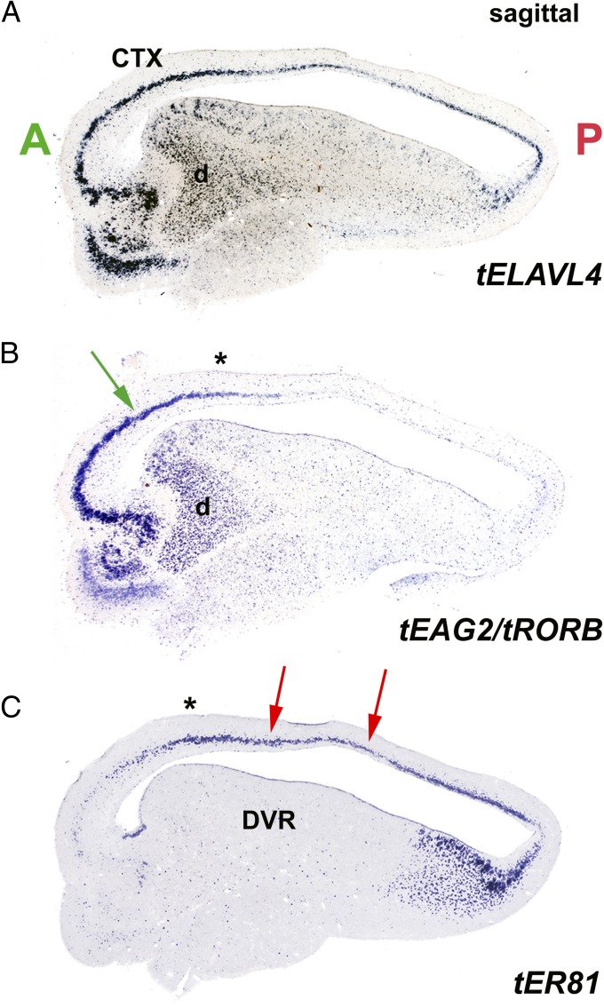
Fig. 5.
L4/I and L5/O cell-type markers identify multiple cerebral cortical fields in the turtle. (A) ELAVL4 ISH demonstrates the pyramidal layer of turtle cortex (CTX) and the neurons of the DVR in a parasagittal cross-section through turtle telencephalon. (B) Two-probe one-color ISH for L4/I markers EAG2 and RORB demonstrates strong expression rostrally in cortical area D2 (green arrow). Labeling in the underlying DVR picks out the thalamorecipient sensory zones of the turtle anterior DVR, including the dorsal area (d) that receives visual input from the nucleus rotundus (4). (C) The L5/O marker ER81 labels intermediate and posterior fields of turtle cortical area D2 (red arrows). (B and C) Asterisks indicate registration between these serially adjoining parasagittal sections, serving to mark the transitional field in area D2 that contains both cell types. A, anterior; P, posterior.

