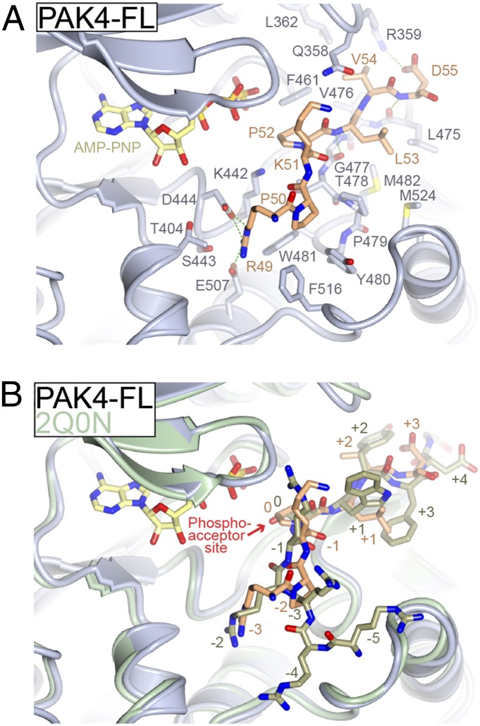
Fig. 4.
Mode of binding for the type II PAK autoinhibitory region. (A) Structural details of pseudosubstrate binding to PAK4 catalytic domain. Structure of PAK4-FL is shown with residues discussed in the text shown in stick format and labeled. Pseudosubstrate is colored orange. Hydrogen bonds are colored green. (B) Comparison of pseudosubstrate binding to a consensus substrate peptide. Crystal structure of PAK4 catalytic domain bound to a consensus substrate sequence (PDB ID code 2Q0N) is shown in green. Labels for substrate and pseudosubstrate indicate number of residues distal from the phosphoacceptor site (labeled and denoted 0).

