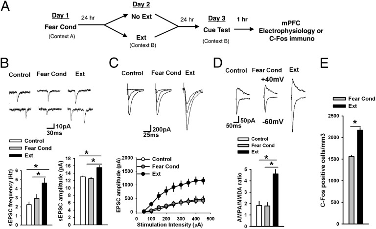
Fig. 2.
Fear extinction modifies glutamatergic synaptic transmission in IL L5 pyramidal neurons of P23 mice. (A) Paradigm design. (B) Frequency and amplitude of sEPSCs in IL L5 pyramidal neurons from control (n = 17 neurons, 6 mice), fear-conditioned (Fear Cond, n = 17 neurons, 6 mice), and fear-extinguished P23 mice (Ext, n = 16 neurons, 6 mice). Both the frequency and amplitude of sEPSCs were significantly increased in fear extinguished mice. (Upper) Examples of sEPSCs. (C) EPSC amplitude in IL L5 pyramidal neurons from control (n = 10 neurons, 5 mice), fear-conditioned (n = 9 neurons, 5 mice), and fear-extinguished P23 mice (n = 11 neurons, 5 mice). EPSC amplitude was significantly increased in fear extinguished mice compared with control group. (Upper) Examples of EPSCs evoked by 100-, 200-, and 300-μA stimulation of L2/3. (D) AMPA/NMDA ratio in IL L5 pyramidal neurons from control (n = 13 neurons, 5 mice), fear-conditioned (n = 8 neurons, 5 mice), and fear-extinguished P23 mice (n = 11 neurons, 5 mice). AMPA/NMDA ratio was significantly increased in fear extinguished mice. (Upper) Examples of EPSCs evoked at −60 and +40 mV by 150-μA stimulation of L2/3. (E) Histograms showing the average density of c-Fos–labeled nuclei in the IL across behavioral treatment group reveal increased density of c-Fos expression in the IL of P23 fear-extinguished mice compared with fear-conditioned mice (P23 Fear Cond 1,557 ± 34.15; P23 Ext 2,169 ± 56.06), Student t test, *P < 0.01. See also Figs. S2–S10.

