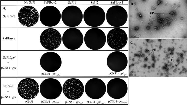
Fig. 1.
Ppi interference with phage 80α. (A) Approximately 108 bacteria were infected with phage 80α, plated on phage bottom agar, and incubated 36–48 h at 32 °C. Plates were stained with 0.1% TTC in TSB and photographed. Genes cloned into pCN51 were induced with 1.0 μM CdCl2. (B and C) Electron microscopic analysis of sedimented particles from (B) phage 80α lysate of RN4220 + pCN51 and from a (C) 80α lysate of RN4220 + pCN51 – ppispb2. CP, complete phage 80α particles; FT, free phage tails; PH, phage proheads.

