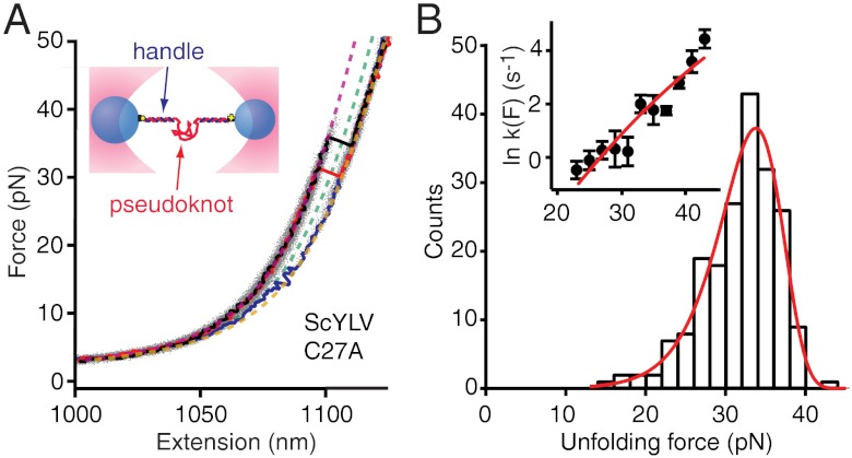
Fig. 1.
Force spectroscopy of ScYLV C27A pseudoknot. (A, Inset) RNA containing the pseudoknot flanked by handle sequences was annealed to DNA strands complementary to the handles and attached to beads held in optical traps. Individual FECs (black, red, blue) are plotted above the aggregated data from 200 FECs (grey). Most FECs (black, red) show a monotonic rise of force with extension up to approximately 30 pN, at which point the extension increases abruptly as the RNA unfolds. A few FECs (blue) unfold at lower forces with a smaller length increase, indicating the RNA started in a different structure composed of fewer nucleotides. WLC fits to the elasticity of the handles, and unfolded RNA, used to determine the contour length change upon unfolding, are shown for three different states of the RNA: fully folded (purple); fully unfolded (brown); incompletely folded (green). (B) The distribution of unfolding forces from the natively folded pseudoknot FECs (black) is well-fit by Eq. 1 (red), yielding parameters describing the mechanical resistance to unfolding. (B, Inset) Unfolding rate as a function of force (black) is well-fit by Eq. 2 (red).

