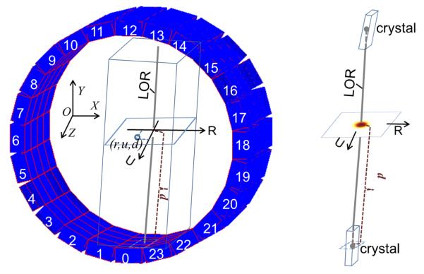
Figure 1.
An illustration of the 3-D LOR-PDF concept. Left: The PET detector ring is shown with image-space and LOR-PDF-space reference frames (x,y,z) and (r,u,d), respectively. The rectangular tube with the LOR as central line is the 3-D volume of the LOR-PDF. The (x,y,z) origin is at the center of the scanner FOV, but is shown offset for clarity. Right: A sample LOR-PDF distribution in one r-u plane.

