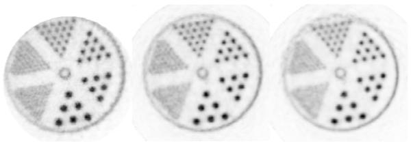
Figure 12.
Comparison of the experimental 18F hot rod in-water phantom reconstructed with the ray-tracing OSEM-2D (left), iso-Gaussian MOLAR (middle), and LOR-PDF MOLAR (right) protocols. The images were summed over 20mm in axial direction, the axial extent of the hot-rod section, to reduce statistical noise effects.

