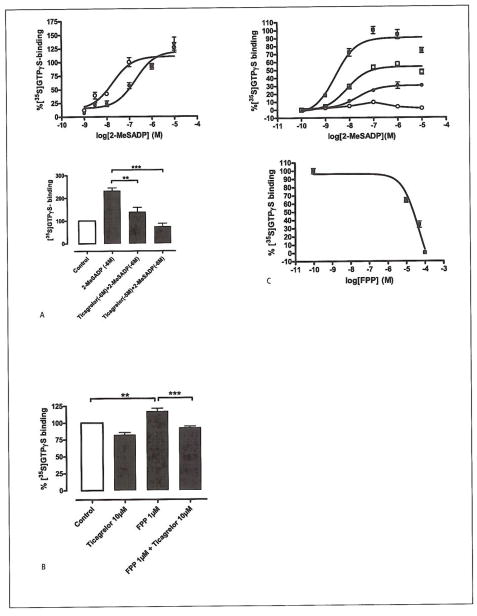
Figure 5. Effects of FFP on ADP-induced [35S]GTPγS binding.
A) Upper panel: 2-MeSADP stimulated [35S]GTPγS binding in isolated platelet membranes. Adding the P2Y12-receptor antagonist ticagrelor, shifted the concentration-response curve to the right. Platelet membranes treated with increasing concentrations of 2-MeSADP (○) display an EC50 value of 17 nM, while in platelet membranes pre-incubated with ticagrelor (60 nM) (●) the EC50 value is increased to 0.4 μM. Lower panel: The bar graphs represents the effect of ticagrelor (1 and 10 μM, respectively) on 2-MeSADP-mediated P2Y12 receptor in platelet membranes (ANOVA: **p<0.01, ***p<0.001, n=4). Untreated platelet membranes where used as control. The number of disintegrations per minute (dpm) from basal and fully stimulated cells (signal-to-noise) in the assay was 3,191 ± 239 and 6,507 ± 324 dpm (mean ± SEM), respectively. B) Bar graph representing the stimulating effect of 1 μM FPP on platelet [35S]GTPγS binding. This effect was suppressed by 10 μM of the P2Y12 receptor antagonist ticagrelor, which indicates that FPP interacts with the P2Y12 receptor. (ANOVA: **p<0.01, ***p<0.001, n=4). Untreated platelet membranes where used as control. C) Upper panel: 2-MeSADP stimulated [35S]GTPγS binding in isolated platelet membranes. Dose response curves were generated in the presence of increasing concentrations of FPP. 2-MeSADP (control), EC50=2.6 nM (■), 2-MeSADP with; [FPP] 10 μM, EC50=7.8 nM (□), [FPP] 50 μM, EC50=22 nM (●), and [FPP] 100 μM, EC50=n.d. (○). Increasing the FPP concentration depressed the maximal signal and shifted the concentration-response curve to the right. The radioactivity counts from basal and fully stimulated cells (signal-to-noise) in the assay were 3,217 ± 70 and 6,727 ± 199 dpm (mean ± SEM), respectively. Lower panel: 2-MeS-ADP-stimulated [35S]GTPS binding in isolated platelet membranes. Platelet membranes were stimulated with 2-MeSADP (1 μM) in the presence of increasing concentrations of FPP. The IC50 value was calculated to be 45 μM.

