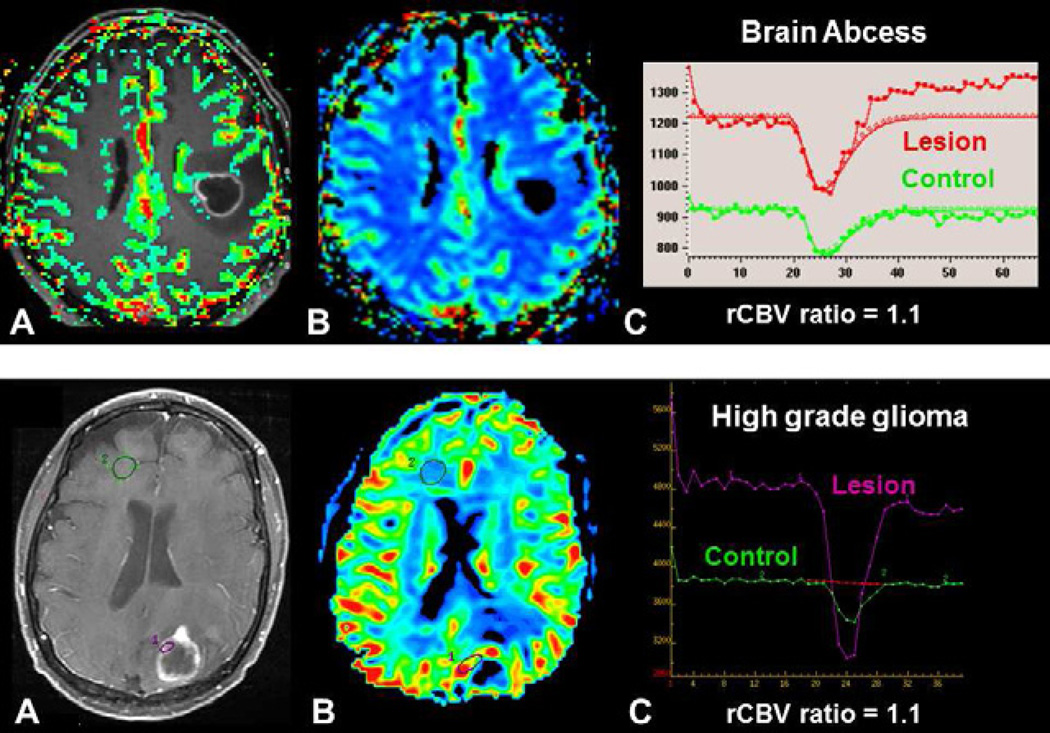
Fig. 2.
Differential diagnostic of brain tumor using DSC-MRI. Both patients presented with a mass demonstraing central necrosis and peripheral enhancement (A, D). In a brain abscess, (upper row) DSC-MRI demonstrates a low rCBV ratio (C) without visible increased perfusion on the rCBV color map (B), whereas in a brain tumor, in this case a metastasis, (lower row), DSC-MRI demonstrates an increase perfusion within enhanced parts of the lesion (E) with a high rCBV ratio (F).

