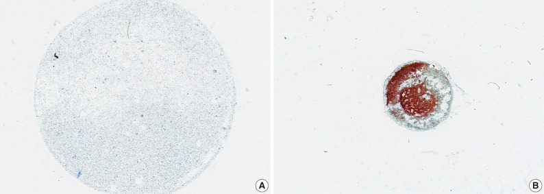
Fig. 1.
Scan image of slides prepared via CellprepPlus® (A) and conventional smear (CS) (B). (A) CellprepPlus® liquid-based cytology has a thin, evenly distributed cell layer with a clean background in a well-defined circular area. (B) CS shows irregular distribution and overlapping of cells in all areas of the slide.

