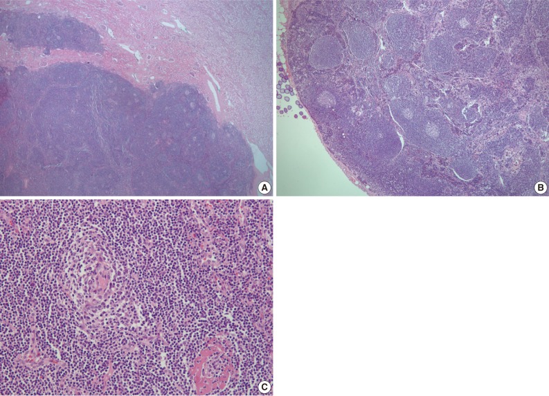
Fig. 3.
Lower power-view shows extensive lymphoid infiltration forming germinal centers in the renal cortex and medulla (A) and several small regressively transformed germinal centers surrounded by a broad mantle zone in the paraaortic lymph node (B). High power-view shows a hyalinized lymphoid follicle and many blood vessels in and around the follicles (C).

