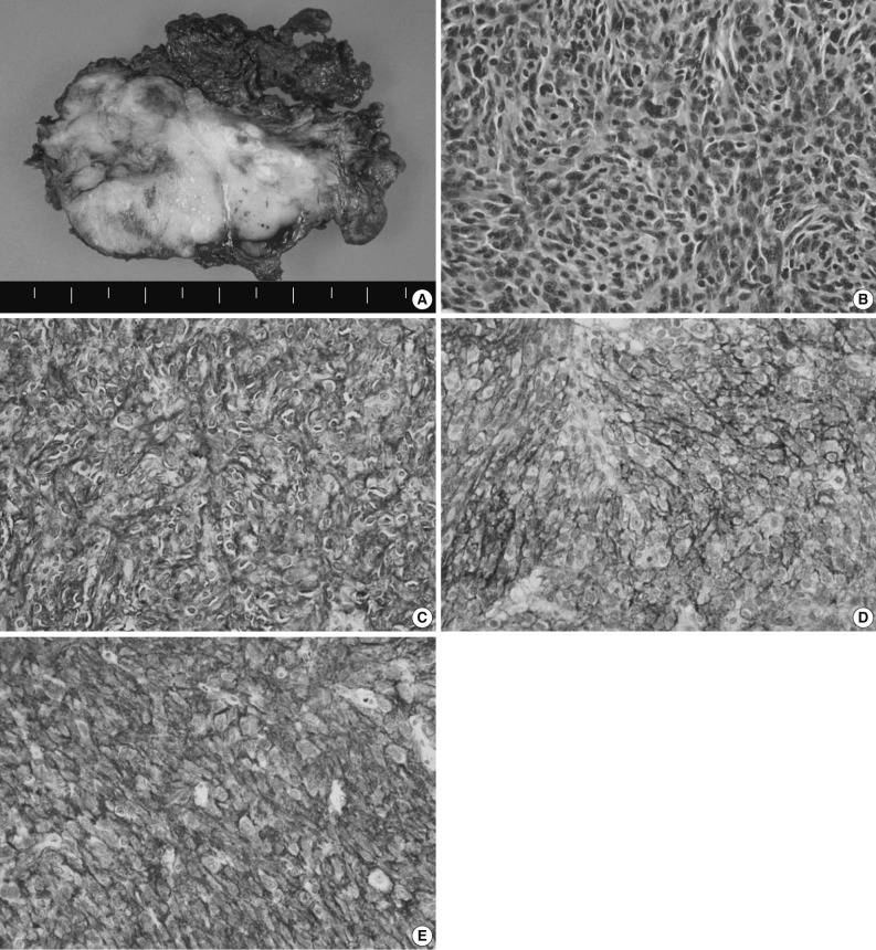
Fig. 3.
Histologic and immunohistochemical findings. (A) Grossly, the mass shows a fibrotic cut surface with a grayish white color. (B) Histopathologically, there is a proliferation of oval to spindle-shaped cells with round, oval or elongated nuclei, vesicular or granular chromatin and an ill-defined margin. (C-E) The immunohistochemical stainings for CD21 (C), CD23 (D), and CD35 (E), markers specific for follicular dendritic cells.

