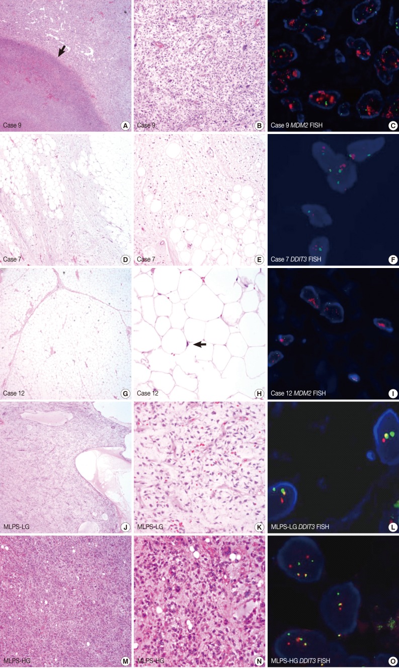
Fig. 1.
(A-C) Case 9. A 49-year-old man with a 40-cm sized retroperitoneal mass. Original diagnosis, myxoid liposarcoma (MLPS); revised diagnosis, dedifferentiated liposarcoma. (A) The low-power view reveals a tumour necrosis (arrow) which causes a myxoid change in the surrounding tumor. (B) In this case, there are pleomorphic tumour cells with high mitotic activity. (C) A high amplification of the murine double minutes (MDM2) gene (red signal) is detected on the fluorescence in situ hybridization (FISH) analysis. (D-F) Case 7. A 47-year-old man with a 4.5-cm sized mass on the chest wall. Original diagnosis, MLPS; revised diagnosis, myxoid lipoma. (D, E) Adipocytes with mild focal atypia are intermingled with fibromyxoid stroma. (F) No rearrangement is identified on the FISH analysis of the DDIT3 gene (a break-apart probe). (G-I) Case 12. A 45-year-old man with a 10.7-cm sized mass in the neck. Original diagnosis, lipoma; revised diagnosis, well-differentiated liposarcoma. (G, H) Mature adipocytes with mild atypical nuclei (arrow). (I) The amplification of the MDM2 gene (red signal) is detected on the FISH analysis. (J-L) Low-grade MLPS (MLPS-LG). (J, K) Loose myxoid tumour showing cystic changes with arborization of the capillary vessels. (L) A break-apart rearrangement of the DDIT3 gene is detected on the FISH analysis. (M-O) High-grade MLPS (MLPS-HG). (M, N) Tumor cells with a round-to-oval shape showing a nuclear pleomorphism in the myxoid stroma. (O) Multiple break-apart rearrangements of the DDIT3 gene are detected on the FISH analysis.

