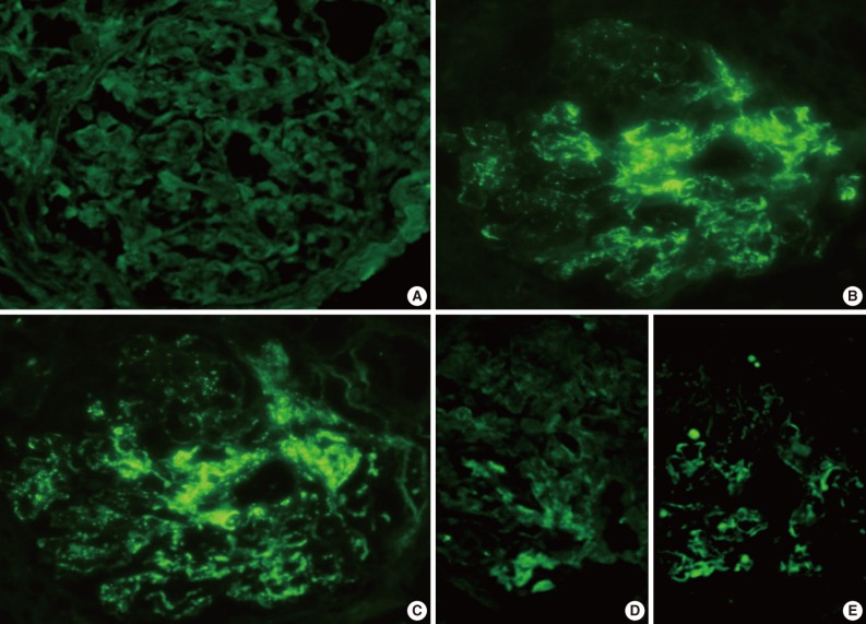
Fig. 2.
Representative photomicrographs of immunofluorescence (IF) staining (patient 2). IF staining for IgG (A) is negative, but there is conspicuous coarsely granular positive IF staining for IgA (B) and C3 (C) along the glomerular capillary wall (3+), and segmentally in the mesangial areas (2+). IF staining for kappa (D) is weakly positive, and lambda (E) shows 1+ segmentally staining in mesangial areas and peripheral capillary walls.

