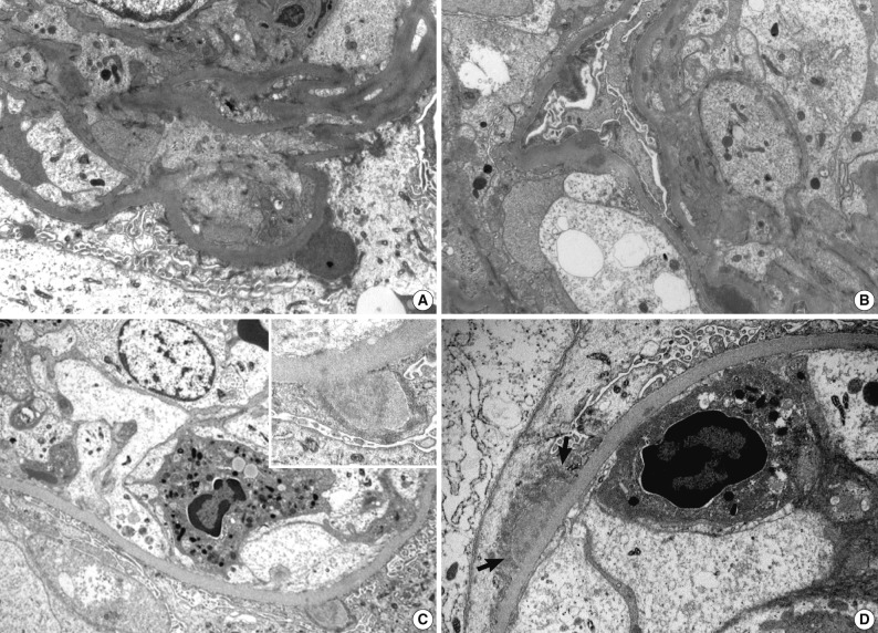
Fig. 3.
Electron microscopic features of two renal biopsies (patients 3 and 5). (A, B) Patient 3 show subepithelial electron dense deposits ('humps'), intramembranous and mesangial deposits. (C) Patient 5 shows a large subepithelial deposit with proliferation of endothelial and mesangial cells, and a neutrophil in the lumen. Partially resorbed subepithelial 'hump' with electron lucency. In (D), there is a broad based subepithelial deposit (arrows) attracting neutrophil in the capillary lumen (A, B, ×8,000; C, ×7,000; inset in C, ×15,000; D, ×12,000).

