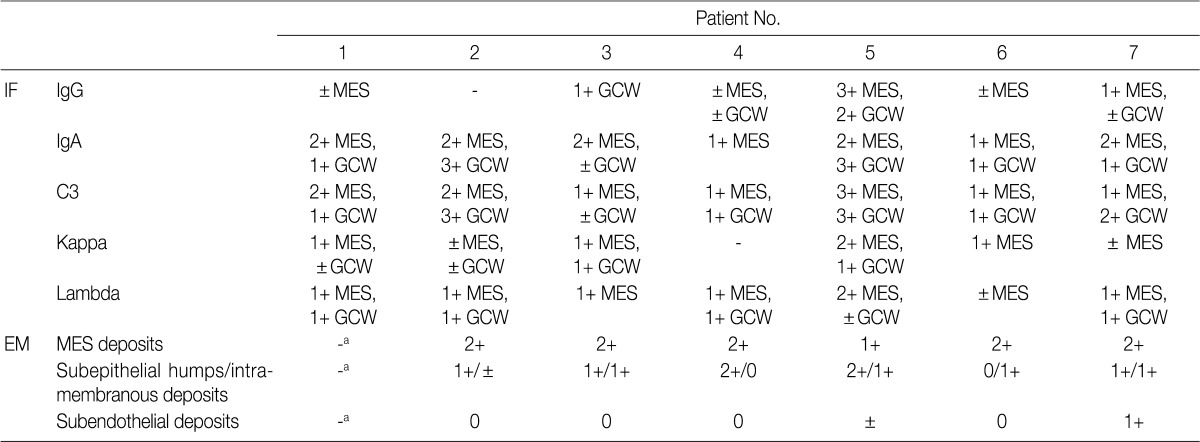
Table 3.
IgA-dominant acute postinfectious glomerulonephritis: immunofluorescence and electron microscopic findings in renal biopsy

Deposits are graded from 0 to 3+ (0, absent; 1+, mild; 2+, moderate; 3+, marked).
IF, immunofluorescence; MES, mesangial; GCW, glomerular capillary wall; EM, electron microscopy.
aNo glomerulus included in EM sample.
