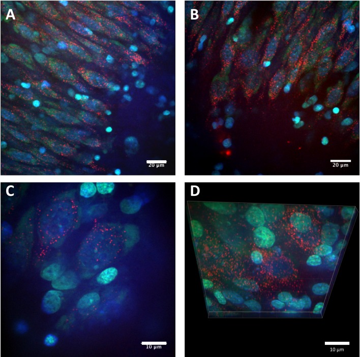
Figure 2. Confocal images of cell uptake of the CL4 QD–Palm1 complex.
(A–D) Representative images of cell bodies located in the CA3 cell layer of the rat hippocampus. QDs (red) containing the CL4 coat were conjugated to Palm1 and applied to the hippocampal slice culture preparation for 24 h. Cells were fixed and imaged with confocal microscopy (Nissl is green, DAPI is blue). The QDs showed neuronal targeting and intracellular localization, exhibiting a punctate, perinuclear distribution. Scale bars in A, B are 20 μm. C, D are 10 μm.

