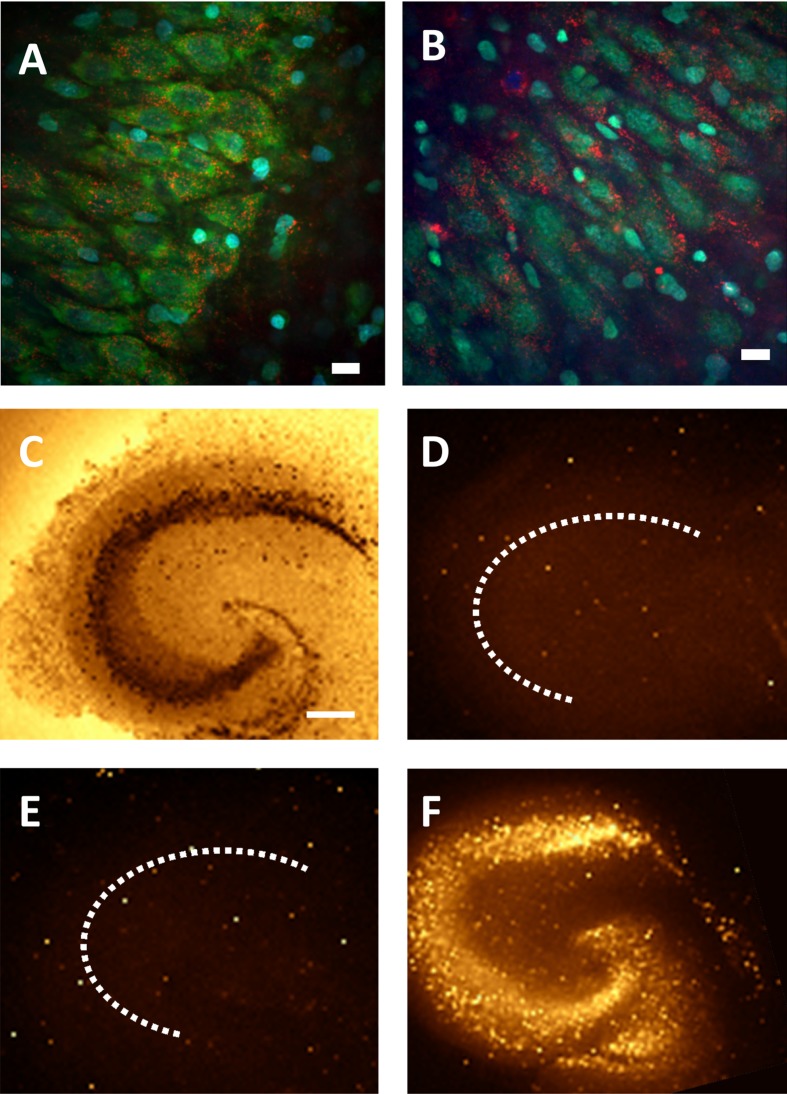
Figure 4. Lack of QD-induced cytotoxicity in rat hippocampal slices after QD–Palm1 treatment for up to 72 h.
CL4 QD–Palm1 (red) after 48 h (A) and 72 h (B) in culture (Nissl is green, DAPI is blue). (C–F) Sytox staining. We observed no QD-induced cytotoxicity to the neurons after 72 h in culture. (C) NeuN stain showing cytoarchitechture. (D) Negative control. (E) CL4 QD–Palm1 after 72 h. (F) Positive control. Scale bars in (A,B) are 20 μm and in (C) are 200 μm.

