
Figure 1.
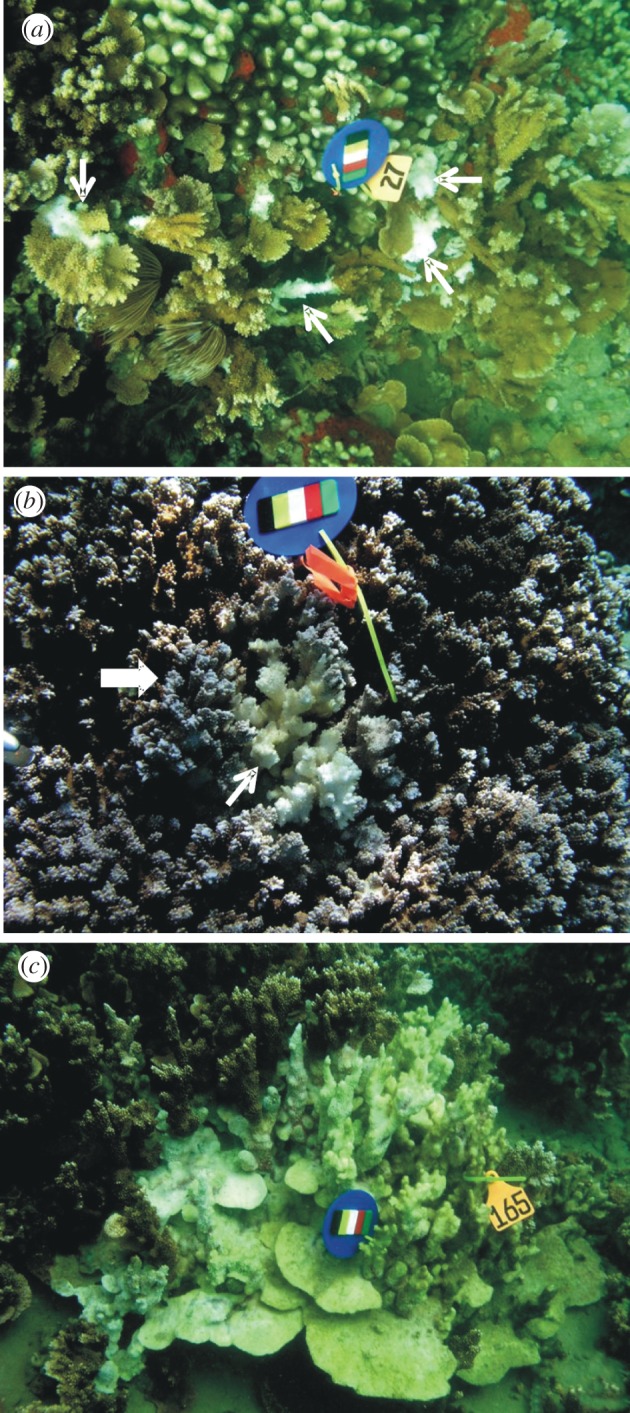
Gross lesions of WS in Montipora capitata. (a) Multifocal; note lesions indicated by white arrows. (b) Locally extensive; note central area of tissue loss revealing bare white skeleton (small white arrow) surrounded by an ill-defined band of pale tissues (block arrow). (c) Diffuse; note extensive area of tissue loss revealing intact bare white skeleton with some green colouring indicating overgrowth of algae. (a–c) Scale bars, 17 cm.