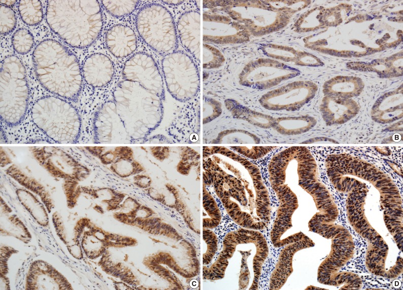
Fig. 1.
Immunohistochemical analysis of PIWIL2 expression in colon cancer and adjacent non-cancer tissue (×200). PIWIL2 protein shows a negative expression in normal glands (A). In colon cancer tissue, PIWIL2 expression shows the cytoplasmic and cytonuclear pattern. The intensity of PIWIL2 expression is graded as weakly positive (B), moderately positive (C), and strongly positive (D).

