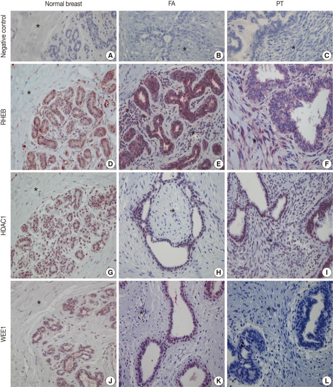
Fig. 1.
Immunohistochemical (IHC) findings. The IHC stains with negative control serum for normal breast tissue (A), fibroadenoma (FA) (B), and phyllodes tumor (PT) (C) are all negative. IHC stains for Ras homolog enriched in brain (RHEB) reveal positive reactions in the nuclei and cytoplasm of epithelial and stromal cells of normal breast (D), FA (E), and PT (F). IHC stains for histone deacetylase1 (HDAC1) are positive in the nuclei of the epithelial and stromal cells of normal breast (G), FA (H), and PT (I). IHC stains are positive for WEE1 homolog (WEE1) in the nuclei of epithelial cells of normal breast (J), FA (K), and PT (L). The areas marked with an asterisk (*) are stromal cells.

