Figure 2.
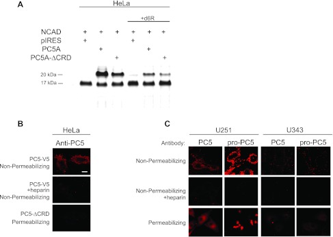
Processing of NCAD by PC5A and immunocytochemical localization of PC5A and its prosegment in U251 and U343 cells. (A) Transfections were done as in Figure 1 except that HeLa cells were transfected with either full-length PC5A (here, we only used the major PC5A isoform throughout [25]) or PC5A-ΔCRD. PC5A with a deleted CRD (PC5A-ΔCRD) cleaved NCAD to a much lesser extent compared to full-length PC5A. (B) Immunocytochemistry was carried out to look at PC5A localization in transfected HeLa cells both under nonpermeabilizing and permeabilizing conditions, as described [27]. PC5A C-terminally tagged with a V5 epitope was cloned in the pIRES2-EGFP vector, and hence, cells were probed with a monoclonal Ab-V5 [27]. Cells were transfected with either full-length PC5A, in the absence or presence of heparin and staining was carried out under nonpermeabilizing conditions to detect cell-surface localization. Cells were also transfected with PC5A-ΔCRD and stained under permeabilizing conditions to identify intracellular localization. (C) Immunocytochemistry of U251 and U343 cells demonstrate localization of endogenous PC5A and its inhibitory prosegment at the cell surface and intracellularly in a perinuclear site. An NT-PC5A Ab detected total PC5A protein, and an Ab against the PC5A prosegment (proPC5A) detected only the prosegment containing proteins [27,50]. Staining was carried out under nonpermeabilizing conditions with or without heparin or under permeabilizing conditions. Bar, 10 µm.

