Figure 1.
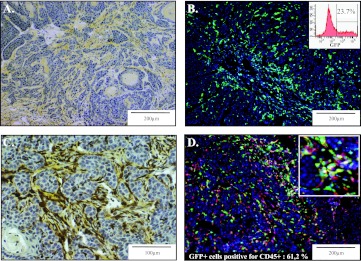
BM-derived cells recruited in the stroma of skin tumors. PDVA malignant keratinocytes were transplanted into mice engrafted with BM from GFP transgenic mice. (A) Intense desmoplastic reaction evidenced within the skin tumor by collagen staining using saffron coloration (yellow). (B) GFP+ BM-derived cells recruited in the tumor. Flow cytometry analysis of cells isolated from the tumor through enzymatic dissociation revealing that 23.7% of cells were GFP+ (insert). (C) Combined GFP immunoperoxidase staining of GFP+ cells (brown) and collagen deposition through saffron coloration (yellow). (D) Detection of GFP positivity (green) and immunostaining for CD45+ inflammatory cells (red). A higher magnification is shown (insert). The percentage of CD45+ cells expressing GFP was assessed by a computerized method of quantification.

