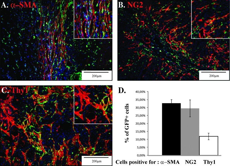
Figure 2.
Characterization of BM-derived fibroblastic cell subsets in the tumor. PDVA malignant keratinocytes were subcutaneously transplanted into both flanks of mice, which have been engrafted with BM derived from GFP transgenic mice. Representative images of cryosections of squamous cell carcinoma tumor tissue infiltrated with GFP+ BM-derived cells (green) and immunostained for the presence of (A) α-SMA, (B) NG2, and (C) Thy1 (red). A higher magnification is shown (insert). (D) Computerized quantification of the colocalization of GFP signal with α-SMA, NG2, and Thy1 immune signals.

