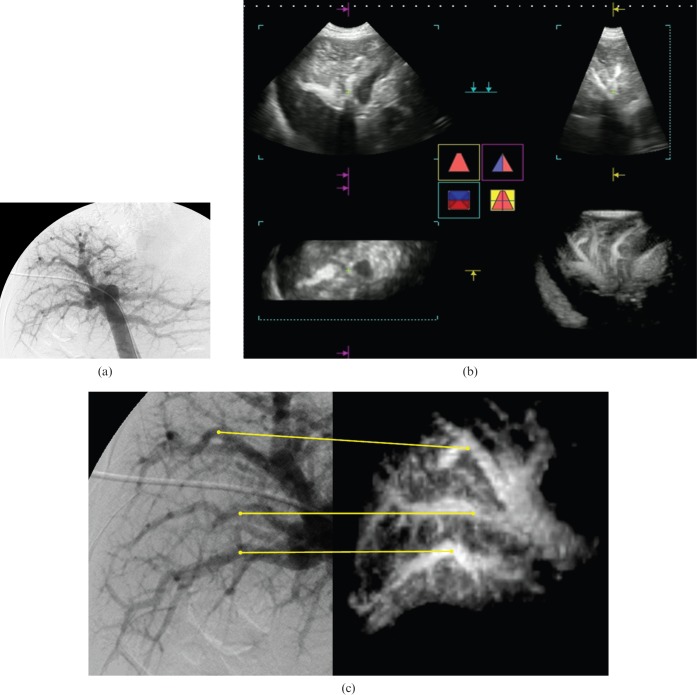
Figure 2.
A 55-year-old male with idiopathic portal hypertension (IPH) (case 9). (a) Portogram obtained by percutaneous transhepatic portography (PTP). This portogram had positive findings of paucity of medium-sized portal branches, irregular and often obtuse-angled division of the peripheral branches, occasional abrupt interruptions of the peripheral branches, an avascular area beneath the liver surface and an increase in the very fine vasculature around the large intrahepatic portal branches with no finding suggestive of cirrhosis, according to the review results by reviewer III. This patient was diagnosed as having IPH by a score of +5 on the PTP image. (b) Multiplanar reconstruction image. Three plane images (upper left, horizontal image; lower left, coronal image; upper right, sagittal image) provided only fragmented information of vascular findings, which were hard to compare with the angiographic images. The lower right image shows dendritically expanded intrahepatic portal vein appearances. This rendered stereoscopic image was rotated to correspond with the angiographic image and used as the contrast-enhanced three-dimensional (3D) ultrasound sonogram. (c) Portograms obtained by PTP and contrast-enhanced 3D ultrasound with Sonazoid® (GF Healthcare, Oslo, Norway). Corresponding portal veins between the PTP image (left side) and the 3D ultrasound image (right side) are indicated by the lines. Contrast-enhanced 3D ultrasound with Sonazoid had positive findings of paucity of medium-sized portal branches, irregular and often obtuse-angled division of the peripheral branches, occasional abrupt interruptions of the peripheral branches and an increase in the very fine vasculature around the large intrahepatic portal branches with no finding suggestive of cirrhosis, according to the review results by Reviewer II. This patient was also diagnosed as having IPH by a score of +4 on the 3D ultrasound image.

