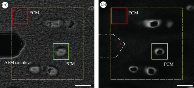
Figure 1.
(a) Phase contrast and (b) fluorescence images illustrating the selection of PCM and ECM testing regions in the deep zone of porcine cartilage as seen during AFM testing. Localization of type VI collagen immediately surrounding cell-sized voids was used to identify the PCM in IF-labelled samples. The AFM cantilever is shown in (a) and outlined in (b), with the red circle indicating the approximate location of the spherical tip. Scale bars, (a,b) 20 µm. (Online version in colour.)

