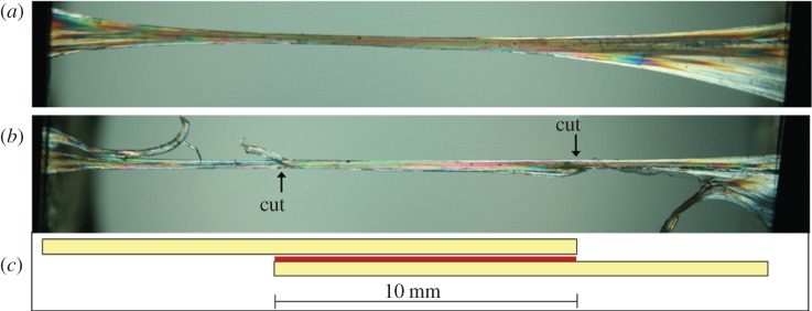
Figure 2.
An illustration of fascicle dissection for testing of the interfascicular membrane. (a) Two intact fascicles bound by fascicular membrane viewed under polarizing light; (b) one end of each fascicle has been cut, leaving a section of intact fascicular membrane of 10 mm length; (c) schematic of fascicle dissection, with fascicles in yellow and interfascicular membrane in red. (Online version in colour.)

