
Figure 2.
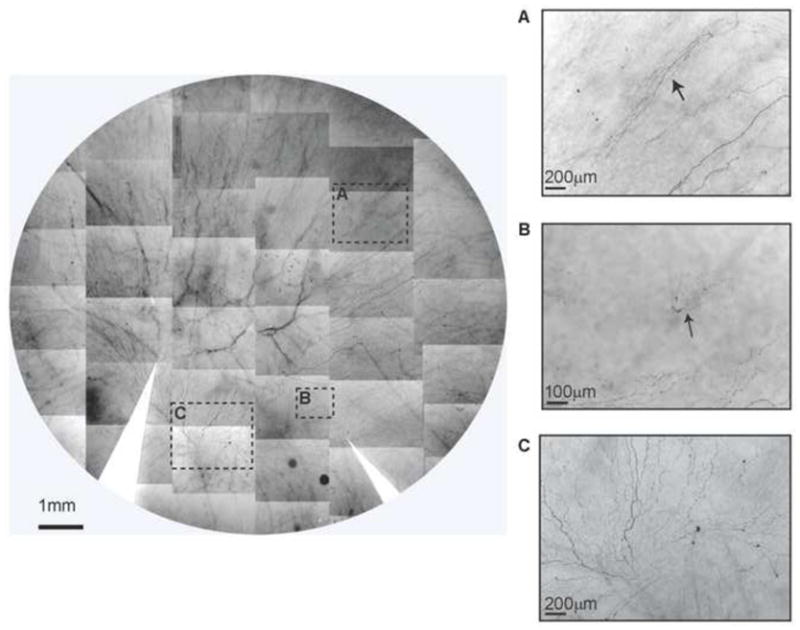
Whole mount view of corneal epithelial nerve distribution in a diabetic cornea after prolonged insulin-dependent diabetes mellitus (IDDM). Images were taken from the left eye of a 58-year-old female with IDDM for 13 years. The montage of images outlined a frame that shows the nerve distribution in detail. The images (A, B) show newly-regenerating epithelial nerve leashes (arrows) with abundant empty spaces without innervations. Image C shows the detailed nerve distribution in the merging area. The long bundles running from the superior quadrants converge in a counter-clock pattern at the inferior quadrant close to the limbal area. Several short nerve bundles, representing newly regenerating nerves, are visible in the lower part.