
Figure 3.
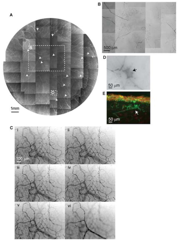
A) Whole mount view of corneal stromal nerve distribution in a diabetic cornea taken from a 43-year-old male with insulin-dependent diabetes mellitus (IDDM) for 5 years. Numerous nerve fiber loops and neuropathic lesions (arrows) are present in the whole cornea. B) Highlighted image shows the nerve fiber loops present in the center. C) Topographic images of the same area show the detailed architecture of corneal nerve loops at different depths. Images were recorded in time-lapse mode from the stroma surface (I) to the middle of the stroma (VI). D) and E) are representative images of diabetic neuropathies in a whole mount and in a transected view.