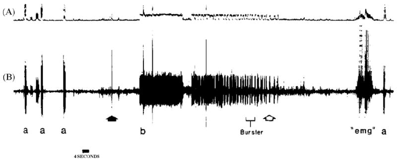
Fig. 4.

Recording from left ulnar nerve at wrist level. A = integrated neurogram; B = simultaneous discriminated neurogram. Symbols: a = stimulation of receptive field in little finger; solid arrow = head flexed to left and extended; b = onset of abnormal unitary bursting, temporally correlating to verbalized report of paresthesias reaching the hand; open arrow = head returned to neutral position; “emg” = voluntary muscle artifact; a = original receptive field reconfirmed.
