Fig. 6.
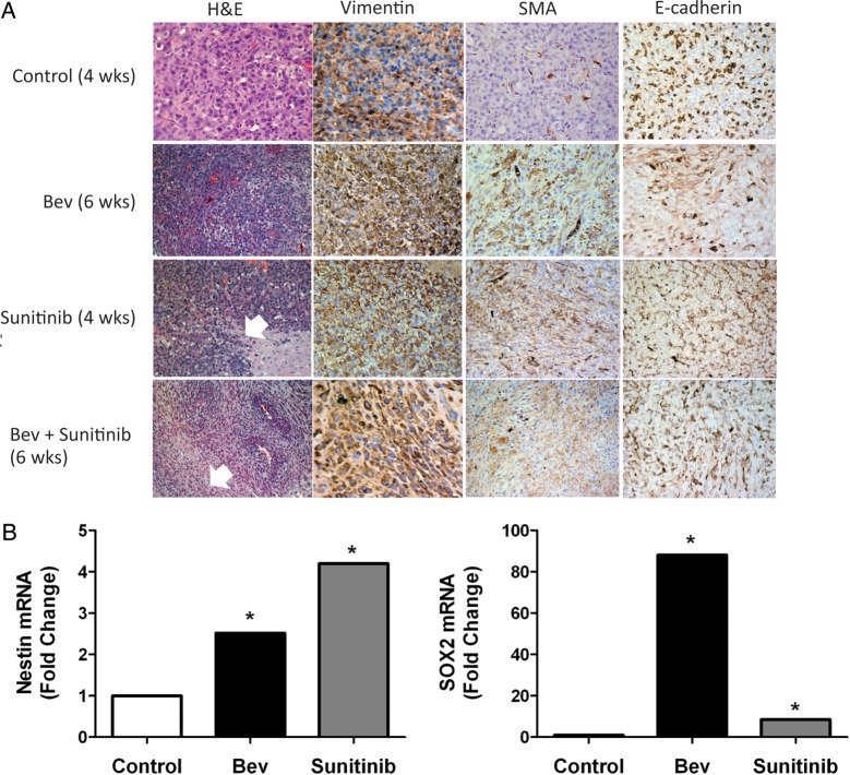
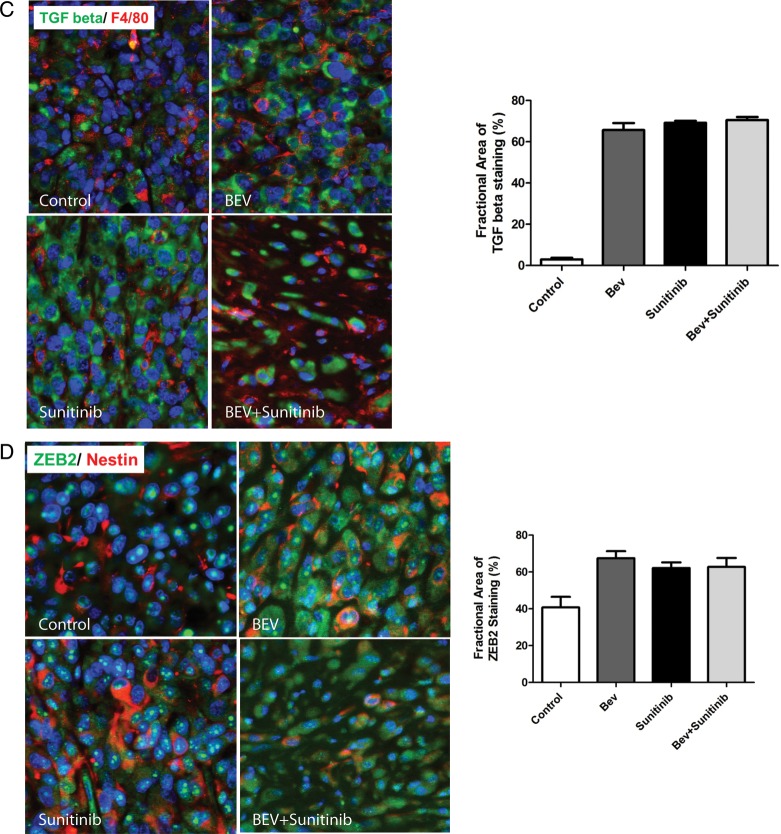
Antiangiogenic therapy induces mesenchymal changes to glioma tumor tissue in vivo. (A) Tumors treated with antiangiogenic therapy induced aggressive (invasive) behavior and histologic characteristics characteristic of mesenchymal tumors (see white arrows). Expression of mesenchymal markers vimentin and smooth muscle actin (SMA) are increased following antiangiogenic therapy, whereas e-cadherin expression decreases. (B) Tumors treated with bevacizumab and sunitinib show a significant increase in the expression of the glioma stem cell markers nestin and SOX2. (C) Expression levels of TGF beta and F4/80 in U87 glioblastoma xenograft tumors in response to antiangiogenic therapy. Tissue slides from xenografts were stained with anti-TGF beta (green) and anti-F4/80 (red) antibodies as described in “Materials and Methods.” The bar graph represents the percentage of TGF beta-positive cells in each condition under ×200. (D) The expression of ZEB2 and nestin in U87 glioblastoma xenograft tumors in response to antiangiogenic therapy. Xenografts tissue was stained with anti-ZEB2 (green) and anti-nestin (red) antibodies as described in “Materials and Methods.” The bar graph represents the percentage of ZEB2-positive cells for each treatment condition under ×200.


