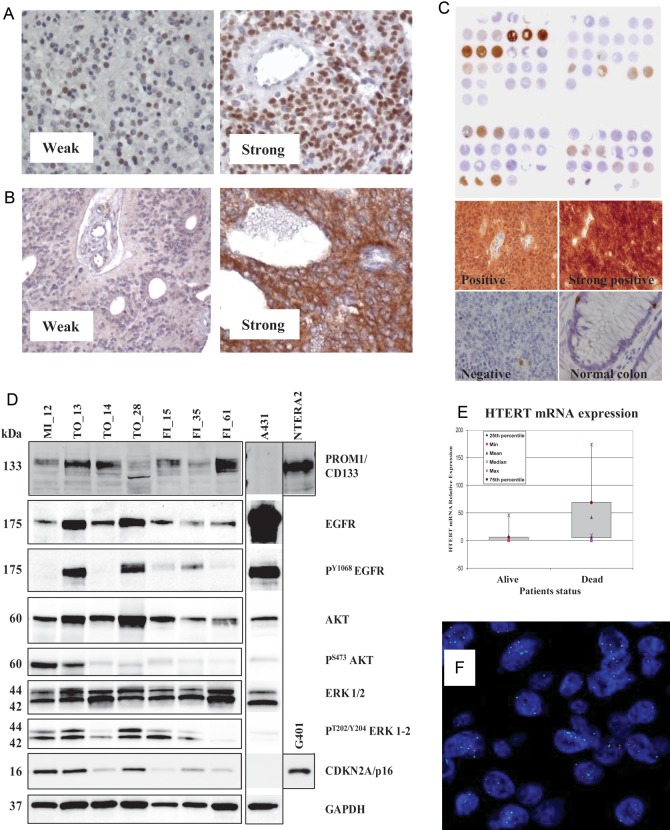
Fig. 1.
Representative examples of IHC stainings of ependymoma samples for (A) nucleolin, (B) EGFR, and (C) CDKN2A/p16 protein expression. (D) Analysis of protein expression by Western blotting from fresh-frozen ependymoma tissues. Equal amounts of protein lysate from cell lines A431, NTERA2, and G401 were loaded as controls. Lanes 2 and 4 correspond to supratentorial ependymomas, while other cases are infratentorial. (E) Differential HTERT mRNA expression in tumor tissue from ependymoma patients who experienced dichotomous outcomes, as detected by qRT-PCR. (F) FISH analysis of an ependymoma sample carrying 1q gain (green signal).

