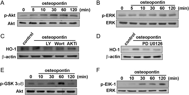
Fig. 4.
Involvement of PI3K/Akt in osteopontin-induced HO-1 expression in U251 glioma cells. Cells were incubated with osteopontin for indicated time periods. The phosphorylation of Akt (A) and ERK (B) were determined by Western blot analysis. (C and D) Cells were pre-incubated with LY294002, wortmannin, Akt inhibitor (AKT i), or PD98059 or U0126 for 30 min followed by stimulation with osteopontin for 24 h, and HO-1 protein expression was analyzed by Western blot. (E and F) Cells were incubated with osteopontin for indicated time periods, and cell lysates were then immunoprecipitated with GSK3- or Elk-1–fusion protein agarose beads. One set of immunoprecipitates was subjected to 10% SDS-PAGE and analyzed by immunoblotting with the anti-phospho-GSK3α/β antibody or anti-phospho-Elk-1 antibody. Equal amounts of the immunoprecipitated kinase complex presented in each kinase assay were confirmed by immunoblotting of Akt or ERK. Results are expressed as 4 independent experiments.

