
Figure 6.
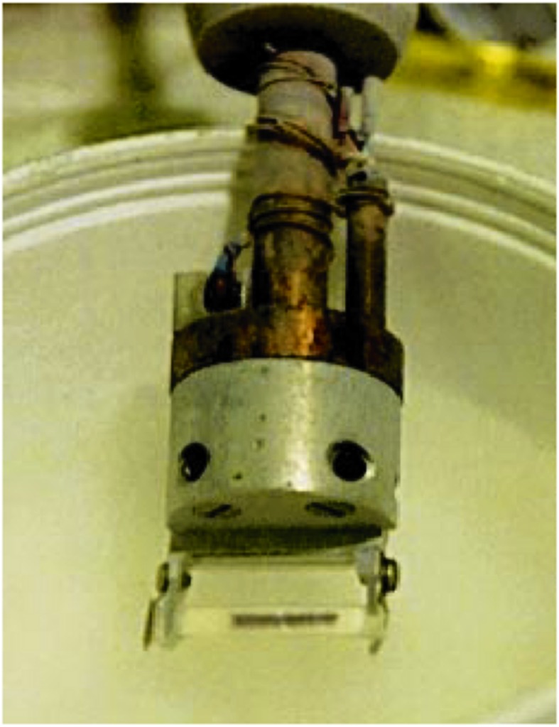
Color centers in a sample of Saccharomyces cerevisiae Cox11, 1 mM Cu(I) (pH 7.2 Bis-Tris-Propane, 20% v/v glycerol) following a 3 h exposure to an X-ray beam at a sample temperature of 10 K in a Cu K-edge XAS experiment (SSRL BL7-3). The profile of the beam on the sample contained in a transparent polycarbonate cuvette can be clearly seen in the lower part of the figure. Note that the apparent horizontal mis-alignment of the beam profile is due to the fact that the sample was oriented at 45° so that on the reverse side the beam is displaced towards the other end of the sample.