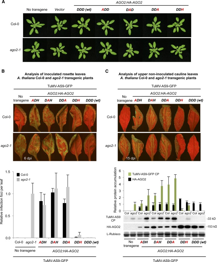
Figure 3.
Phenotypic and Molecular Analyses of Col-0 and ago-2-1 T3 Transgenic Plants Expressing Wild-Type or Modified AGO2 Forms.
(A) Pictures of 21-d-old Col-0 (top panel) and ago2-1 (bottom panel) T3 transgenic plants.
(B) Analysis of TuMV-GFP-AS9 viral infection in inoculated rosette leaves at 6 d after inoculation. Top, pictures were taken at 6 d after inoculation under UV light. Bottom, the number of infection foci for TuMV-AS9-GFP was expressed relative to those in ago2-1 (12 ± 3 foci per leaf). The graph shows the average and sd for 32 leaves and eight plants per treatment.
(C) Analysis of viral infection in upper noninoculated cauline leaves at 15 d after inoculation. Top, pictures were taken at 15 d after inoculation under UV light. Bottom, accumulation of TuMV-AS9-GFP CP and HA-AGO2 in cauline leaves from ago2-1 and Col-0 transgenic lines at 15 d after inoculation. Mean (n = 4) relative to TuMV-AS9-GFP CP (green) and HA-AGO2 (black) levels + sd [ago2-1 and DDD (wt) = 1.0 for CP and HA-AGO2, respectively]. l-Rubisco (ribulose-1,5-bisphosphate carboxylase/oxygenase) blot is shown as loading control.

