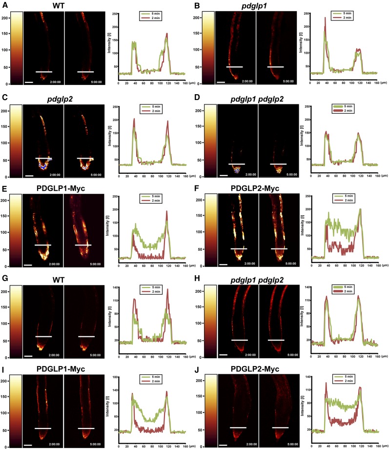
Figure 7.
Symplasmic Permeability Is Enhanced in the Root Tips of Transgenic PDGLP1-Myc and PDGLP2-Myc Plants.
A brief (10-s) application of CFDA (60 µg/mL) to the primary ([A] to [F]) or lateral ([G] to [J]) root tips was used to load CF into the cytoplasm of Arabidopsis root cap and epidermal cells. CF distribution within the root tip was analyzed by confocal microscopy at 2 and 5 min after CFDA application. In wild-type (WT) (A), pdglp1 (B), pdglp2 (C), and pdglp1 pdglp2 (D) primary root tips, CF was restricted to the outer layer of cells. For the PDGLP1-Myc (E) and PDGLP2-Myc (F) seedlings, CF was detected in the central region of the primary root tip within 5 min of CFDA application. For wild-type (G) and pdglp1 pdglp2 (H) lateral root tips, CF was restricted to the outer layer of cells. By contrast, in the PDGLP1-Myc (I) and PDGLP2-Myc (J) seedlings, CF was detected in the central region of the lateral root tip within 5 min of CFDA application. Right panels present quantification of the fluorescence intensity measurements made at the locations indicated by the white bars on the individual roots. Red and green traces represent fluorescence intensity profiles collected at 2 and 5 min after CFDA application, respectively. Color bars on the left side of the confocal image indicate relative fluorescence intensity. Bars = 60 µm.
[See online article for color version of this figure.]

