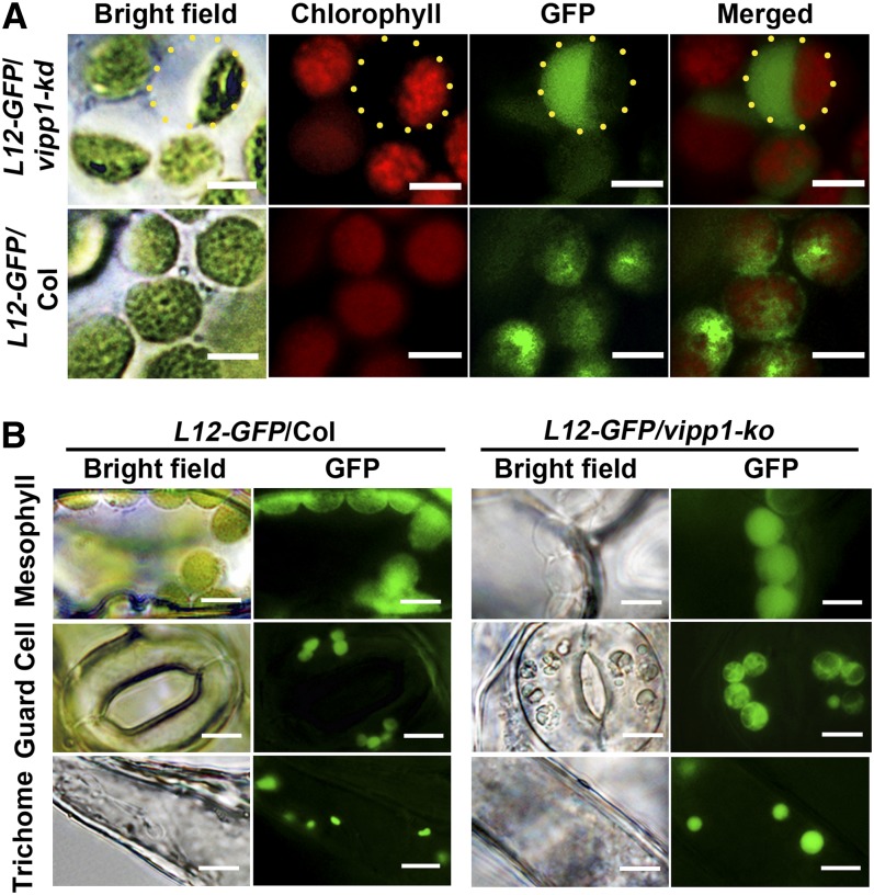
Figure 2.
Swelling of Chloroplasts and Plastids in vipp1 Mutants Visualized Using Stroma-Localized L12-GFP.
(A) Distribution of L12-GFP within chloroplasts. Transgenic lines expressing L12-GFP in Col (L12-GFP/Col) or vipp1-kd (L12-GFP/vipp1-kd) were generated, and their mesophyll chloroplasts were observed by microscopy. Bright-field images and their fluorescent signals corresponding to chlorophyll (red) and GFP (green) are indicated along with merged images. Yellow-dotted circles represent the area of swollen stroma, which is transparent in bright field but detectable by L12-GFP. Bars = 5 µm.
(B) Spherical and enlarged plastids detected in different cell types of the transgenic line expressing L12-GFP in vipp1-ko (L12-GFP/vipp1-ko). Chloroplasts/plastids from mesophyll cells, guard cells, and trichomes were visualized using L12-GFP. Bright-field images (left) and GFP signals (right) are shown. Bars = 10 µm.

