Figure 4.
Chloroplasts and Plastids in vipp1 Mutants Examined Using TEM.
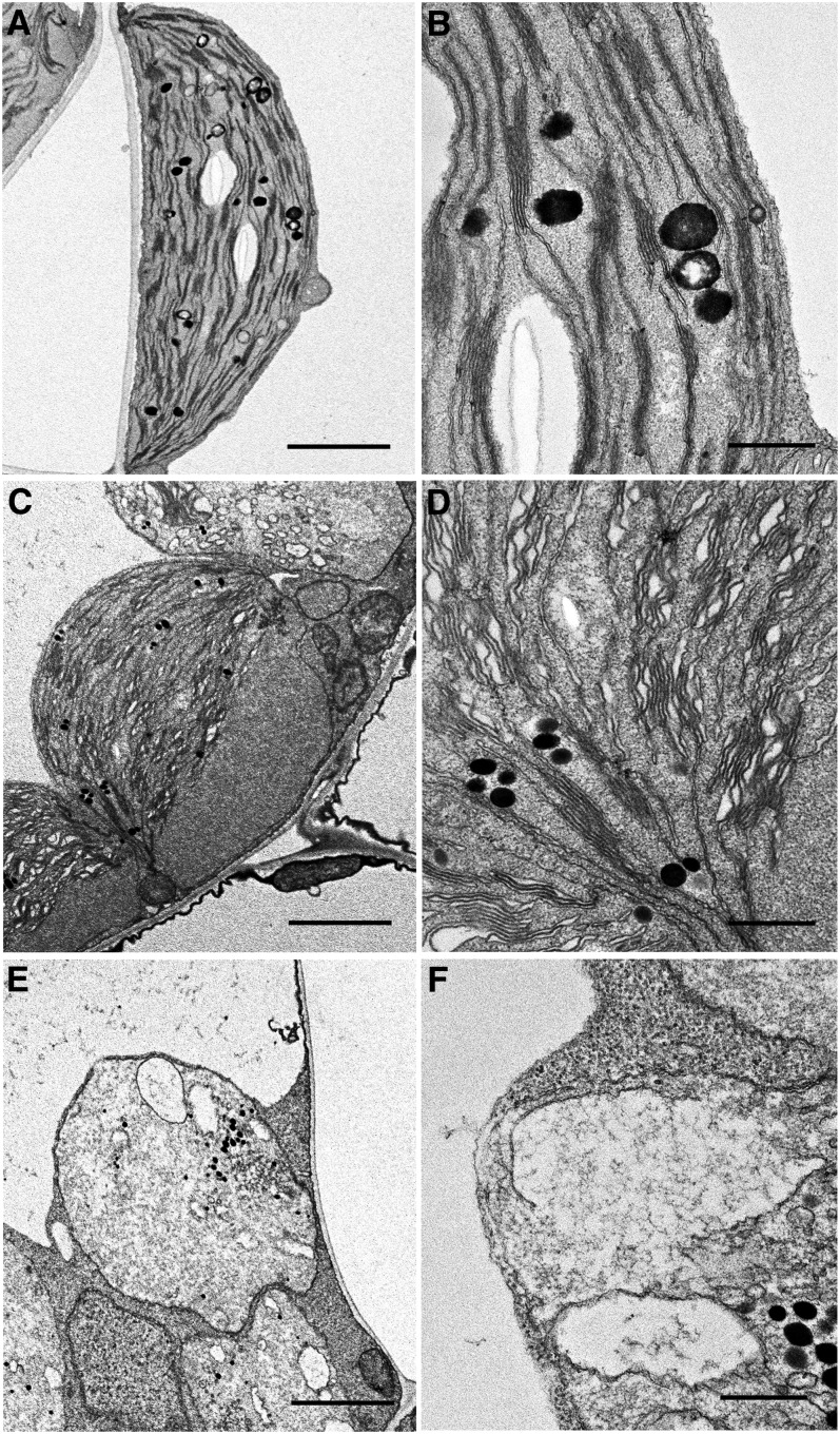
Chloroplast ultrastructure of Col ([A] and [B]), vipp1-kd ([C] and [D]), and vipp1-ko ([E] and [F]) seedlings was observed using TEM. Spherical chloroplasts with extra stromal space, detected in unfixed tissues, are also detected using electron microscopy in vipp1 mutants ([C] and [E]). Magnified electron micrographs corresponding to grana thylakoids show irregular granal stacks in vipp1-kd (D) and vacuolated membrane structures in vipp1-ko (F). Bars = 2.5 µm in (A), (C), and (E) and 500 nm in (B), (D), and (F).

