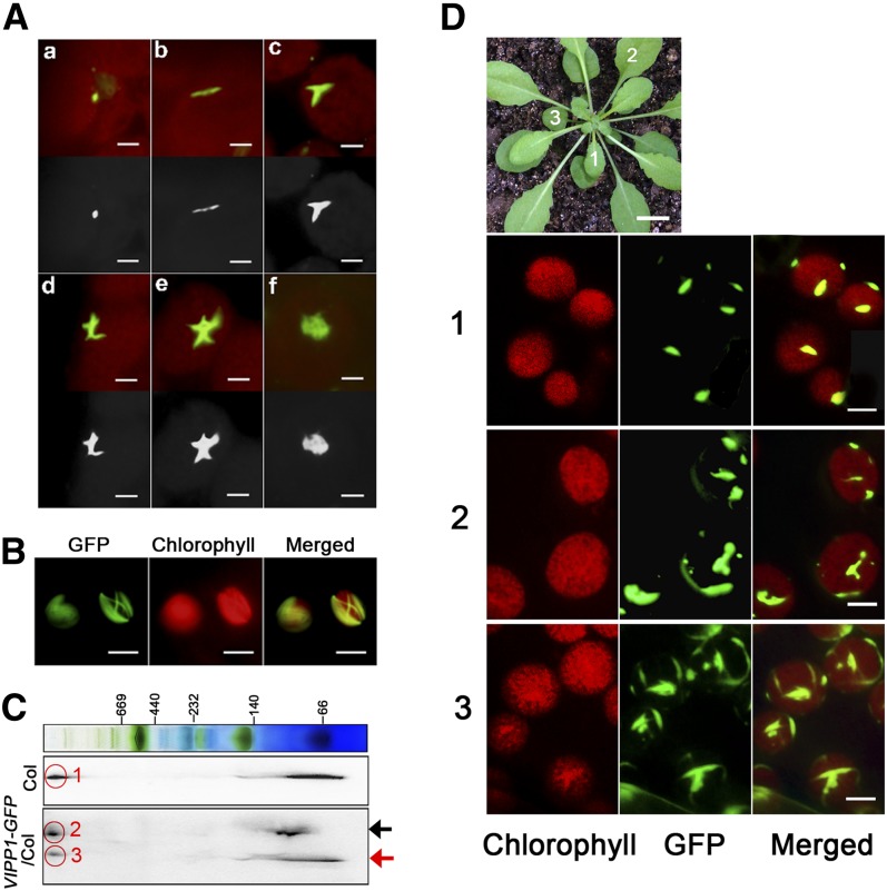
Figure 6.
Microscopy Observation of VIPP1-GFP Reveals Various Supercomplexes along the Chloroplast Envelope.
(A) Images of VIPP1-GFP supercomplexes in chloroplasts of VIPP1-GFP/vipp-kd protoplasts photographed using fluorescence microscopy: Various morphologies, such as a dot (a), line (b), fork (c), cross (d), five-point star (e), and web (f) were detected. Top panels show the original images obtained from microscopy observation, and bottom panels show their black and white images converted by Photoshop software. Bars = 2.5 µm.
(B) Lattice-like structures of VIPP1-GFP occasionally formed along the chloroplast envelope. Bars = 10 µm.
(C) Supercomplex of VIPP1 and VIPP1-GFP in Col and VIPP1-GFP/Col, respectively, monitored using Blue Native-SDS-PAGE. Total chloroplast proteins of Col and VIPP1-GFP/Col were separated by Blue Native-SDS-PAGE and probed with antibodies against VIPP1. Black and red arrows indicate positions of VIPP1-GFP and VIPP1, respectively. The signals within the red circles respectively correspond to the supercomplex of VIPP1 in Col (1), of VIPP1-GFP in VIPP1-GFP/Col (2), and of VIPP1 in VIPP1-GFP/Col (3).
(D) Morphologies of VIPP1-GFP at different stages of leaf development. Top, photograph of a VIPP1-GFP/vipp1-kd plant (bar = 1.0 cm). Leaves marked with numbers (1, 2, and 3) in this plant were subjected to microscopy observation. Chloroplasts from each leaf were observed to detect chlorophyll autofluorescence (left) and GFP (middle). Merged images of both signals are shown on the right panels. Bars = 5.0 µm.

