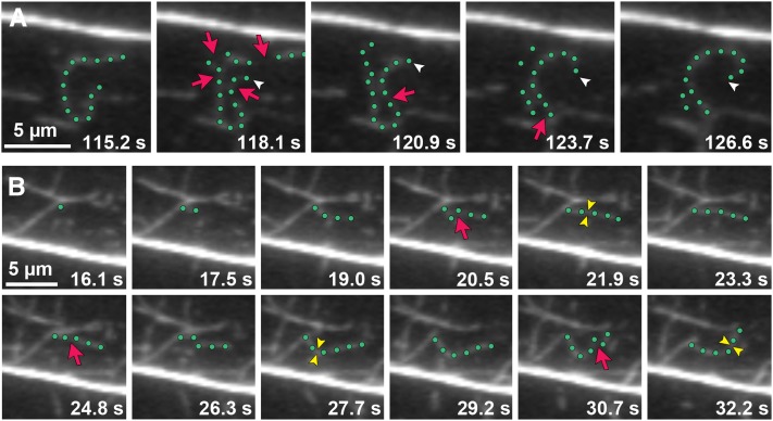
Figure 5.
The Dynamic Behavior of Actin Filament Ends in Epidermal Cells Can Be Tracked.
(A) Time-lapse VAEM series shows an example of an actin filament elongating from a newly created barbed end. The highlighted filament (green dots) stops growing and is severed (red arrows) into several fragments. One filament end (white arrowheads) resumes growth within 5 s after severing (see Supplemental Movie 4 online).
(B) New filaments can be constructed by filament-filament annealing of severed fragments. The highlighted growing filament gets severed (red arrows). Newly created ends join together within ∼1.5 s to form a new filament (yellow arrowheads). Three annealing events occur in the sequential time-lapse frames (see Supplemental Movie 5 online).
Images were taken by time-lapse VAEM from cpb-1 epidermal cells. Bars = 5 µm.

