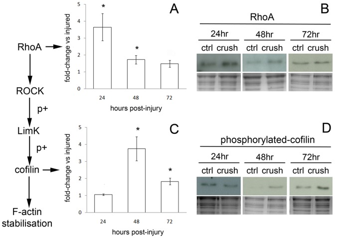
Figure 3. RhoA and phosphorylated cofilin expression are modulated in axoplasm-enriched samples from injured optic nerve.
Injured optic nerves and control contralateral nerves were collected at 24, 48 and 72 hrs and axoplasm-enriched samples generated. Immunoblots for RhoA and phosphorylated cofilin are shown, with coomassie stained gels to determine loading (B&D). Quantification is presented as fold-change vs injured and represent 3 independent experiments ±SEM (A&C); one-way ANOVA plus Tukey post hoc test was used to assess significance, and where indicated (*) equals p<0.05. Immunoblotting for RhoA protein revealed a significant increase at 24 hrs, which persisted up to 48 hrs following injury (A&B). Following the increase in RhoA we observed a lag of 24 hrs before a significant increase in phosphorylated cofilin was detected that persisted until 72 hrs post-injury (C&D).

