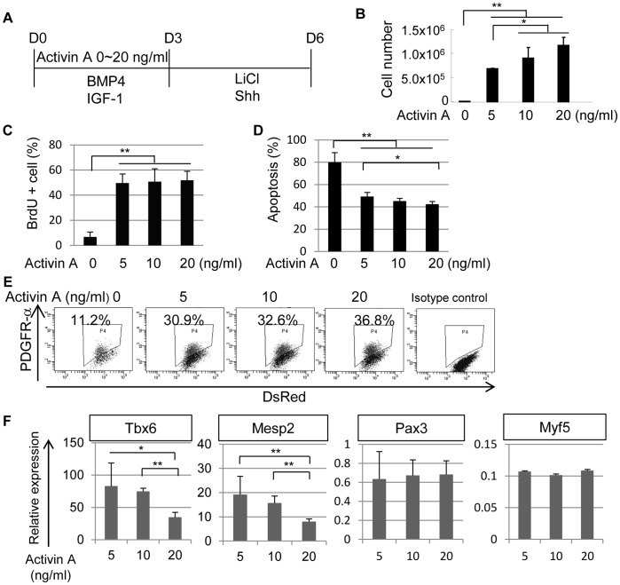
Figure 1. Effects of Activin A on paraxial mesodermal differentiation of mouse iPS cells.
(A) A scheme for paraxial mesodermal differentiation of mouse iPS cells with different concentrations of Activin A from day 0 (D0) to day 3 (D3). Activin A was administrated from D0 to D3 at a concentration of 0–20 ng/ml. The cultures also contained BMP4 (10 ng/ml), IGF-1 (10 ng/ml), LiCl (5 mM), and Shh (10 ng/ml). The cells were analyzed on day 6 (D6) in (B), (E), and (F), or on day 1 (D1) in (C) and (D). (B) Total number of mouse iPS cells after differentiation with the protocol shown in (A) (n = 3). (C) Proliferation of differentiated mouse iPS cells on D1 assessed by BrdU assay (n = 3). (D) Apoptosis of differentiated mouse iPS cells on D1 assessed by a proportion of Propidium Iodide (PI) positive/AnnexinV positive cell (n = 3). (E) Dose-dependent induction of PDGFR-α by Activin A in mouse iPS cell differentiation culture. The percentage indicates the proportion of PDGFR-α+ cells (n = 3). (F) Gene expression profiles of PDGFR-α+ cells in Activin A-induced cultures (n = 3). The expression level of Tbx6 and Mesp2 genes was reduced in a dose-dependent manner. *p<0.05, **p<0.01 between selected two samples.

