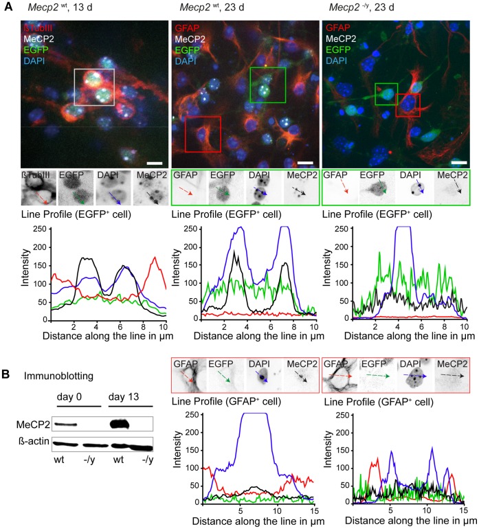
Figure 2. MeCP2 becomes detectable in heterochromatic chromocenters at late differentiation stages.
A. Mecp2 wt and Mecp2 −/y tEG cells were differentiated, fixed, and immunostained with antibodies to MeCP2. Tau promoter driven EGFP expression highlights neuronal cells. Additional markers as beta tubulin III (neuronal cell) and GFAP (astroglia) allow discrimination of different cell populations. In immunofluorescence experiments MeCP2 protein is detected as of day 13 (left) and remains constant thereafter (middle) in EGFP+ cells. Line profiles across chromocenters highlight MeCP2 protein accumulation at these structures (black line) as well as their intense DAPI signal (blue line). Some GFAP+ astroglia revealed a relative weaker MeCP2 signal (black line) compared to neurons. Mecp2 deficient cells (right) were used as control. Bar: 10 µm. B. Immunoblot experiments demonstrate the increase of MeCP2 protein over time in wild type cells (wt) while Mecp2 deficient cells (−/y) show no signal. Beta actin was used as a control for equal loading.

