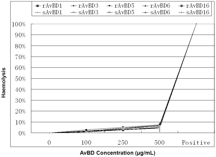
Figure 6. Hemolytic activities of duck avian β-defensins (AvBDs).
Freshly isolated duck red blood cells were incubated with different concentrations of AvBDs (0–500 µg/mL). Release of hemoglobin, as a measure of hemolysis, was measured at 405 nm. Release of hemoglobin upon addition of 1% Triton X-100 was set at 100%. The percentage of hemolysis was calculated as [(A 405 nm, peptide – A 405 nm, PBS)/(A 405 nm, 1% Triton X-100– A 405 nm, PBS)] × 100%. All assays were performed in three independent experiments, with three replicates per experiment, and each point is the mean ± SD. The data were analyzed using SAS [24].

