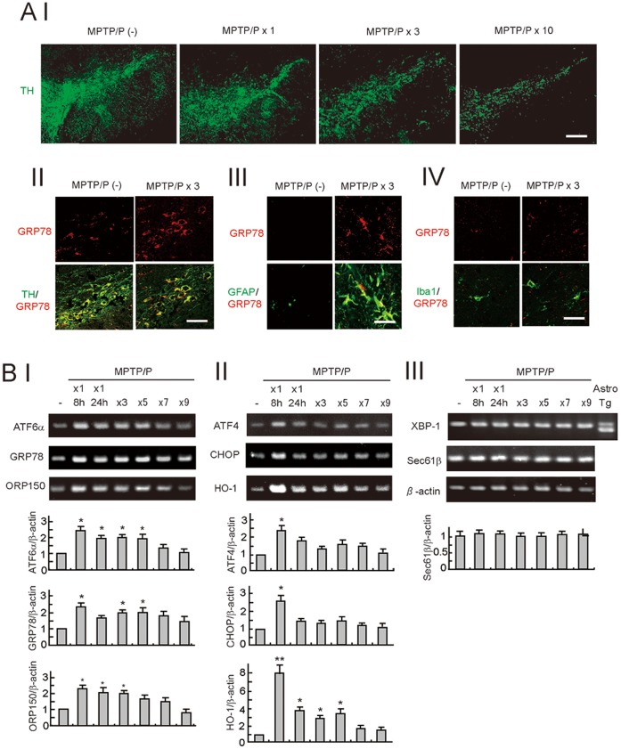
Figure 1. The unfolded protein response (UPR) in a mouse model of chronic MPTP/P injection.
A, Neurodegeneration (I) and UPR activation (II, III, IV) in the SNpc after MPTP/P injections. Brain sections, including the SN from wild-type mice injected with or without MPTP/P were immunostained with the TH, GRP78, GFAP, and Iba1 antibodies. Scale bars = 50 µm (I), 30 µm (II), 20 µm (III), 20 µm (IV). B, Gene expression in the UPR branches after MPTP/P injections. Total RNA (1 µg) isolated from the ventral midbrain of mice was subjected to RT-PCR with specific primers for ATF6α-target genes (I), ATF4-target genes (II), XBP1-target genes, and β-actin (III). The far right lane in (III) indicates the unspliced and spliced form of the XBP1 from cultured astrocytes treated with thapsigargin (an ER stressor). The relative intensity of the bands derived from the mice without MPTP/P injection is designated as one. Values shown are the mean ± S.D. *P<0.05, **P<0.01 compared with mice without MPTP/P administration (n = 4).

