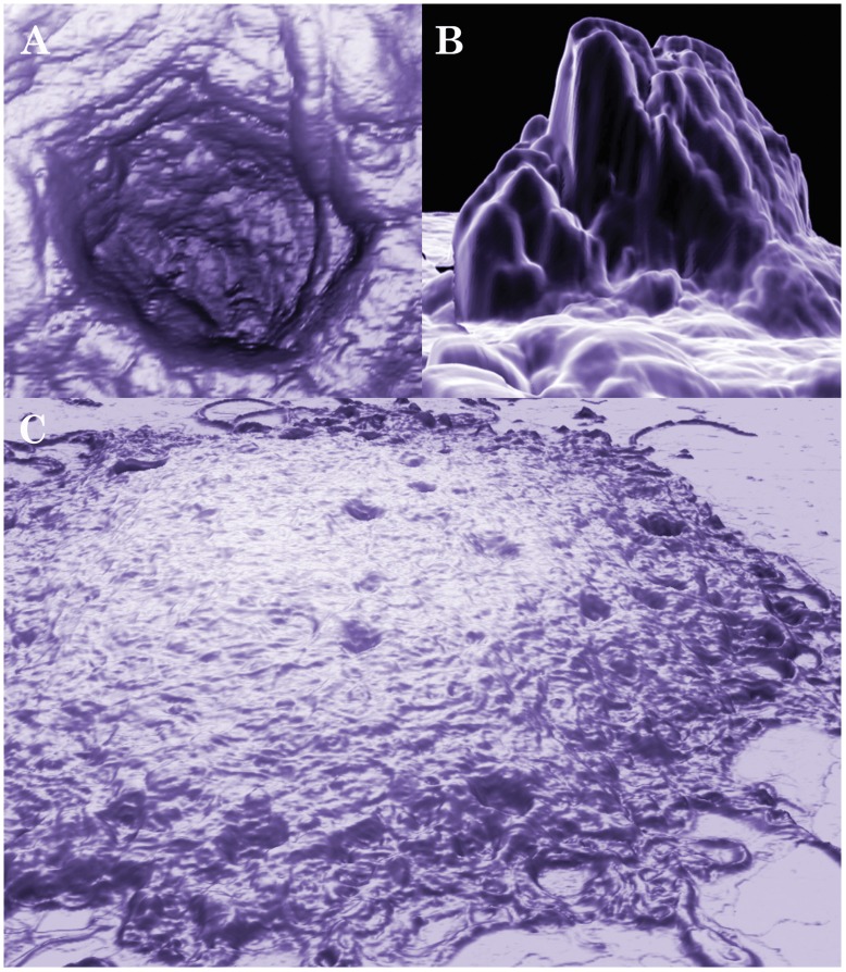
Figure 3. Three-dimensional AFM images of a mature aggregate of Borrelia burgdorferi B31 strain after 20 days.
The preparation of Borrelia burgdorferi cells on mica is described in the Materials and Methods. The scan was conducted with 0.4 Hz using contact mode. A and B show a pit and a protrusion, respectively, of a large mature aggregate as depicted in C. Images A and C were produced with NanoRule© software; image B was produced with a custom meshing utility and MeshLab open-source software.

