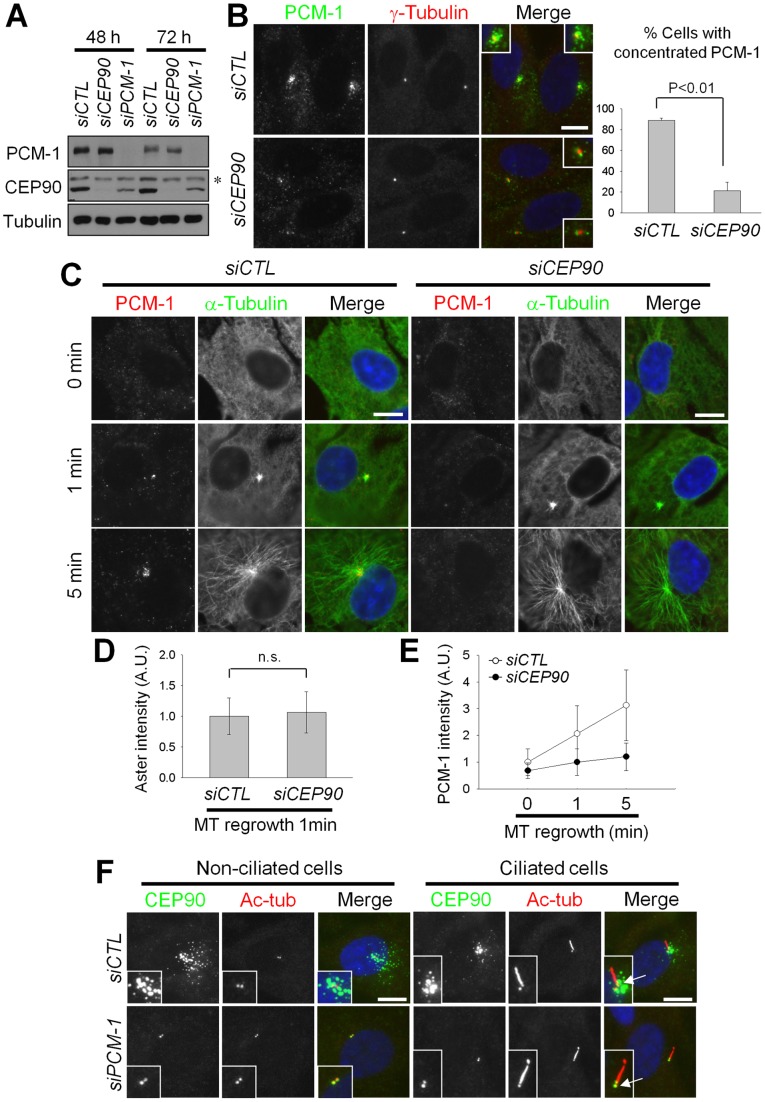
Figure 1. CEP90 is required for the accumulation of PCM-1 granules at the centrosome.
(A) RPE-1 cells were transfected with siCTL, siCEP90 or siPCM-1 and cultured for 48 or 72 h. The cells were subjected to immunoblot analysis with antibodies specific to PCM-1, CEP90 and β-tubulin. An asterisk indicates a non-specific band. (B) The CEP90-depleted RPE-1 cells were co-immunostained with antibodies specific to PCM-1 and γ-tubulin. The resulting images were merged, along with DAPI (nuclear) staining. The insets are magnified views of the centrosomes. The number of cells with centrosome-concentrated PCM-1 was counted. Over 300 cells per experimental group were analyzed in 3 independent experiments. (C–E) The CEP90-depleted RPE-1 cells were cultured in the presence of nocodazole (1 μg/ml) for 3 h and placed in fresh medium for 0, 1 or 5 min. (C) The cells were co-immunostained with the antibodies specific to PCM-1 and α-tubulin. (D) The centrosomal α-tubulin intensities were determined at 1 min in fresh medium. Forty cells per experimental group were analyzed by densitometry in two independent experiments. n.s. indicates not significant. (E) The intensities of centrosomal PCM-1 were determined at the indicated time points. (F) CEP90 localization in PCM-1-depleted RPE-1 cells. The cells were co-immunostained with antibodies specific to CEP90 and acetylated tubulin (Ac-tub). Arrows indicate basal bodies at the base of cilia. The graphs show the mean values and standard errors (B) or standard deviations (D, E). Scale bar, 10 μm.

