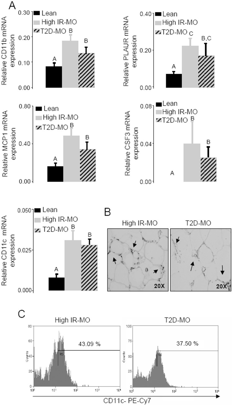
Figure 2. Elevation of infiltrated macrophages in visceral adipose tissue from high IR-MO and T2D-MO subjects.
(A) The mRNA expression of macrophage markers CD11b, PLAUR, MCP1, CSF3 and CD11c was increased in the MO group compared to healthy leans (Duncan; p<0.05). More importantly, the expression of macrophage markers was not increased in the visceral adipose tissue of T2D-MO compared to high IR-MO patients. (B) Immunohistochemical detection of CD68+ macrophages in VAT from both, high IR-MO and T2D-MO patients, characteristically showed macrophages surrounding adipocytes forming the typical crowns. No changes were detected between both morbidly obese groups. Significant differences (Duncan; p<0.05) are indicated with different words. (C) CD11c detection in ATMs through flow cytometry indicated that there was no significance difference in CD11c protein expression in high IR-MO and T2D-MO subjects.

