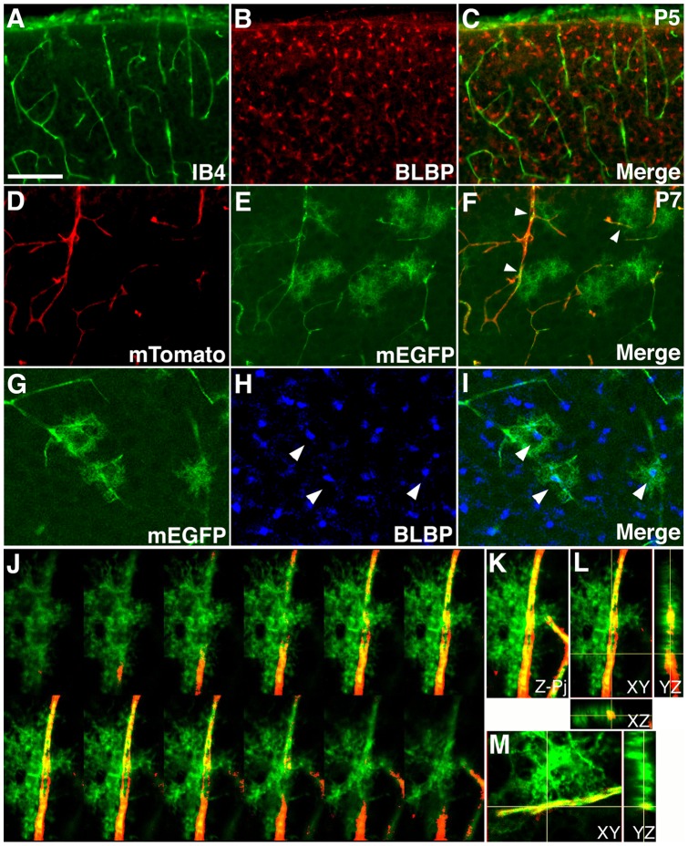
Figure 1. Close interactions between astrocytic processes and blood vessels in the early postnatal mouse cortex.
(A–C) Limited interactions between the cell body of young astrocytes and blood vessels in the mouse cortex at P5. Blood vessels were stained using isolectin B4 (IB4, green in A & C), while young astrocytes were labeled using anti-BLBP antibodies (red in B & C). Limited overlap was observed between IB4 and BLBP staining. (D–F) Close interactions between astrocytic processes and blood vessel in the mouse cortex at P7. An mTomato/mEGFP reporter line was used to labeled astrocytes by nestin-creER mediated recombination induced at E18.0. A sporadic number of astrocyes and their processes were labeled by expression of mEGFP (green in E & F), while blood vessels continued to express a strong level of mTomato (red in D & F). The vast majorities of labeled astrocytes contact blood vessels though their processes (arrowheads in F). Also note green autofluorescence in blood vessels, which does not stain positive for anti-GFP antibodies. (G–I) Confocal microscopy analysis of mEGFP (green in G & I) and BLBP (blue in H & I) staining of brain sections as in (D–F). Each of the mEGFP positive cells overlaps with a single BLBP positive cell body (arrowheads in H & I), indicating that mEGFP positive cells are astrocytes. (J–L) Interactions between astrocytic processes and blood vessels examined by 3-D confocal reconstruction. Astrocytic processed are labeled by mEGFP (green). Blood vessels are labeled by mTomato (red). An astrocyte approaches from the left side and interact extensively with a blood vessel, with some processes wrapping around the vessel and interacting from the opposite side. A montage of selected Z-images is shown in (J), Z-projection in (K), and XY, XZ, and YZ cross-sections in (L). (M) Cross-sections of 3-D reconstruction of a second astrocyte that show clear interactions of individual astrocytic endfeet with a blood vessel. See supplemental information for movies of the 3-D reconstruction. Scale bar in (A), 200 μm for (A–C) and 100 for μm (D–I).

