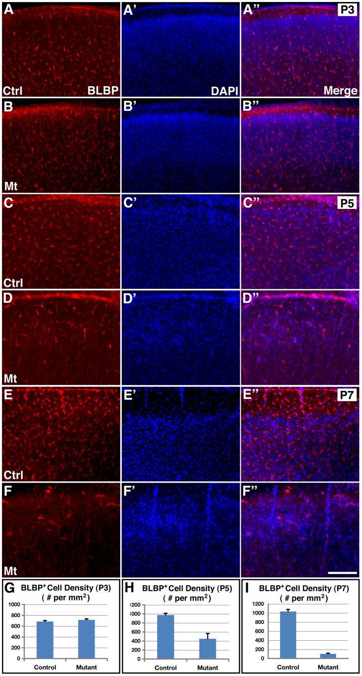
Figure 2. Inhibition of cortical astrogliogenesis by hGFAP-cre mediated orc3 gene deletion in class II mutants.
(A–F”) Young astrocytes were stained using anti-BLBP antibodies (red in A–F and A”–F”), and nuclei were labeled by DAPI staining (blue in A'–F' and A”–F”) in control and orc3 mutant cortices at P3 (A–B”), P5 (C–D”) and P7 (E–F”). No obvious differences were observed in the density of BLBP positive astrocytes between control (A–A”) and mutant (B–B”) cortices at P3. However, obvious reductions were observed in mutants (D–D”) as compared to controls (C–C”) at P5. Young astrocytes also appeared more elongated in the mutant cortex at P5. By P7, BLBP positive astrocytes were almost completely eliminated from mutants (D–D”) as compared to controls (C–C”). (G–I) Quantification of BLBP positive cell density in P3 (G), P5 (H), and P7 (I) control and mutant cortices. Statistical analysis by Student's t test showed no significant changes in mutants at P3 (control, 685.6±19.1/mm2; mutant, 716.0±18.8/mm2; P = 0.27, n = 9), but significant decreases in BLBP positive cell density at both P5 (control, 974.8±38.6/mm2; mutant, 447.7±117.4/mm2; P = 0.001, n = 11) and P7 (control, 1033±47/mm2; mutant, 701±14/mm2; P = 0.0001, n = 4). Scale bar in (F”), 200 μm for (A–F).

